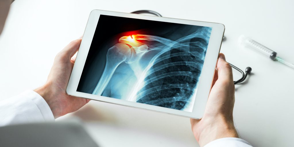
Digital Xray

Digital X-ray, also known as digital radiography, is a modern imaging technique that uses digital sensors to capture images of the internal structures of the body. Unlike traditional film-based X-rays, digital X-rays provide instant images that can be easily viewed, enhanced, and stored electronically. This technology is widely used in medical diagnostics to detect and […]

What is it?

Digital X-ray is a diagnostic imaging procedure that uses digital detectors to capture detailed images of the body’s internal structures. These images are generated using X-ray radiation, which passes through the body and is captured by digital sensors. The resulting images are then processed and displayed on a computer screen, allowing healthcare providers to examine bones, tissues, and organs for abnormalities.

What happens during the procedure:

During a digital X-ray procedure, the following steps typically occur:

  1. Preparation: The patient may be asked to remove any metal objects, such as jewelry, that could interfere with the imaging. Depending on the area being examined, the patient may need to change into a hospital gown.
  2. Positioning: The patient is positioned appropriately, either standing, sitting, or lying down, depending on the part of the body being X-rayed. The radiologic technologist ensures the correct alignment and positioning.
  3. Imaging: The digital X-ray machine is positioned over the area to be examined. The patient is asked to remain still, and sometimes to hold their breath, while the image is taken. The digital sensors capture the X-ray beams that pass through the body.
  4. Image Processing: The captured image is immediately processed and displayed on a computer screen. The radiologist or technologist reviews the image to ensure it is clear and covers the necessary area.
  5. Completion: The patient can typically resume normal activities immediately after the procedure unless otherwise advised by their healthcare provider.

Benefits:

  • High-Quality Images: Produces clear, detailed images that can be easily enhanced and manipulated for better diagnosis.
  • Instant Results: Provides immediate image availability, reducing the wait time for diagnosis and treatment planning.
  • Lower Radiation Dose: Often requires a lower dose of radiation compared to traditional film X-rays.
  • Electronic Storage: Enables easy storage, retrieval, and sharing of images in electronic health records (EHRs).
  • Environmentally Friendly: Eliminates the need for film and chemical processing, reducing environmental impact.

Things to keep in mind:

  • Radiation Exposure: Although the radiation dose is low, it is still important to limit unnecessary exposure. Inform your healthcare provider if you are pregnant or suspect you might be.
  • Motion Artifacts: Remaining still during the X-ray is crucial to avoid blurry images that might require a repeat scan.
  • Preparation Instructions: Follow any specific instructions given by the healthcare provider, such as fasting or wearing loose clothing.
  • Communication: Inform the technologist about any medical conditions or previous surgeries that might affect the X-ray results.
  • Contrast Agents: Some X-rays may require the use of contrast agents to enhance image clarity. Inform the provider of any allergies to contrast materials.

Alternatives:

  • Computed Tomography (CT) Scan: Provides more detailed cross-sectional images of the body using a series of X-rays and computer processing.
  • Magnetic Resonance Imaging (MRI): Uses magnetic fields and radio waves to produce detailed images of organs and tissues without radiation.
  • Ultrasound: Uses high-frequency sound waves to create images of soft tissues and organs, commonly used for abdominal, pelvic, and vascular imaging.
  • Traditional Film X-ray: An older method that uses photographic film to capture X-ray images, though less common with the advent of digital technology.
  • Nuclear Medicine Scans: Involves the use of small amounts of radioactive materials to diagnose and treat various conditions, providing functional information about organs and tissues.